Thursday 11 July 2013

How to make a Glass Brain in Brain Voyager v. 2.2

I realize that it has been some time since I made my last blog post, and many things have happened since April...

I am still in the process of writing up my IFJ manuscript. Having finally sent a first draft to my supervisor, I am currently re-working my figures. This takes A LOT of time!

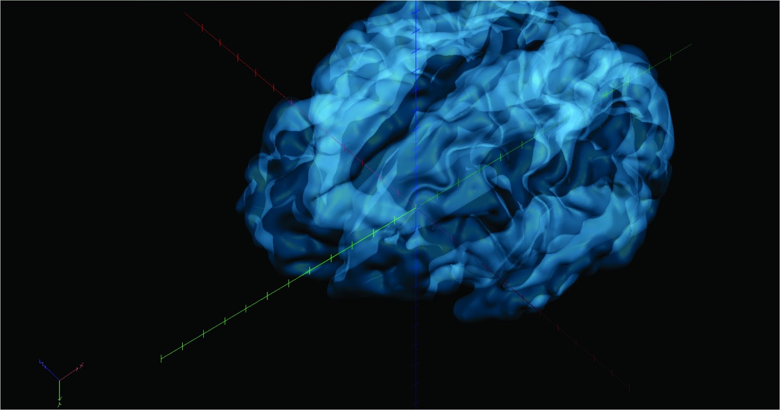
While creating my figures initially I ran in to some problems figuring out how to create a "Glass Brain" image in Brain Voyager from an anatomical scan in which I anatomically landmarked my region of interest. If it took me days to perfect, naturally there must be other people out there with the same inquiry, so i have decided to share the process of creating a figure such as this one...

Step 1

Open Brain Voyager File OpenVMR file (aligned to ACPC, in Talairach coordinates)


Step 2

Open 3D volume tools (little cube on left hand side) Click on Segmentation TabAuto.Segm.

Under segmentation steps, click every box
Under processing space info, should be in talairach
Under cortex reconstruction, click all boxes
Press GO
The BV window for white/grey matter will pop up with a predetermined number. You can choose to change this, however I have left it with the value that Brain Voyager determined → click ok

Let this load. Don't click on anything on the screen (sometimes the mesh will not go to completion if you do). This process should take a couple minutes, depending on the speed of your computer. 

A second tab should be created now, with your original file name + _RH_GM.srf

You will only see one hemisphere, however the left hemisphere was also created, and should be in the same file as your .vmr 


Step 3

Click on the Meshes tab in Brain VoyagerAdd MeshAdd the other hemisphere (the file that has _RH_GM.srf OR _LH_GM.srf). You will now have two 3D hemispheres on your screen


Step 4

Click on the Scene tab Scene overviewThis shows you which meshes you have open + which ones are active. 

Click on the Scene tab Merge Meshes in SceneThis will merge the two hemispheres in to one mesh (much easier to work with when moving through steps 5 and 6)


Step 5

Click on the Meshes Tab → Rendering Options →  Quality display effects →  Click Transparency

OPTIONAL: You can change the degree of transparency under the alpha


Step 6

You can change the color of 3D brain by going to...

Meshes  →  Background and curvature colors  Default and convex color  Choose color here


Adding an anatomically landmarked region of interest

If you have anatomically landmarked a region of interest you can created a mesh version of this region and add it to your glass brain as follows

Step 1

On your .vmr screen, press CTRL+R. This should bring up the volume of interest analysis screen

Select your regions of interest (SHOW VOI)

Under the Options tab, lower right corner of volume of interest analysis GUI, which will bring up a VOI Analysis Options GUI

Click on the VOI Functions Tab


Step 2

Click CREATE under VOI -> Surface Clusters


Step 3

Under Meshes, Click Mesh Morphing, Choose your parameters, Press GO. You have now created morphed mesh regions of interest.


Step 3

Under Meshes, click Add Mesh. Add your Merged.srt brain


Step 4

Finally, make yourself a glass brain while leaving the region of interest opaque 


Tuesday 16 April 2013

New Semester, New Focus

Heading in to the third term of my Masters, I am more prepared for what lies ahead... my entire Masters thesis! And hopefully some more blog posts!

I am now better equipped to analyze functional MRI scans via Brain Voyager and FSL, semi-SPSS literate (thank you Discovering Statistics Using SPSS), getting in to some minor computer programming, and know more tricks than I ever thought I'd know in Microsoft Office Excel.

Having gained all of this necessary skill, I have begun planning the next few months of my life, because I have every intention to finish my Masters on time, with a defense scheduled for May of 2014.

I am finally done working with the IFJ data, will hopefully have something to present at SFN in November, and am working on a manuscript as we speak!

While working on this project, I am also slowly accumulating knowledge on the topic(s) of my Masters thesis: motor learning, Parkinson's disease, visualization, the neurophysiology of dance and NOW... EEG!

I am hoping to incorporate EEG in to my Masters project - I'll update you when I figure out exactly how I am going to do this....

For now, I'm off to read... a lot


Thursday 29 November 2012

Figuring out a research project


The first few months of grad school were definitely overwhelming. A mish-mash of coursework, lab work, T.A-ing, and learning new computer software took up the majority of my time. In the midst of it all, I couldn't help but feel a lingering paranoia that I had not come up with a thesis project, and thus I spent the last two weeks brainstorming a written research proposal for my supervisor. 

The Multisensory Systems Neuroscience laboratory is currently involved with the National Ballet of Canada, working with the dancers in the apprenticeship program to learn more about the neural mechanisms behind motor learning. For my research project, I have decided to investigate the differences between this expert group of dancers and a group of adult non-dancers taking ballet for the first time. Motor learning will be monitored over an 8 month period via fMRI. I am also hoping to examine whether the observed changes in brain activity can be maintained without actually keeping up with the dance classes. I am very interested in Dance Therapy for Parkinson's Disease, and in the long run, I am hoping to relate my research findings to this area, ideally assisting in the optimization of dance therapy for Parkinson's patients. More on the details of my research endeavors to come...

While doing some background reading on my thesis topic, I came across a very interesting article titled Partially Overlapping Neural Networks for Real and Imagined Hand Movements, written by Emmanuel Gerardin, Angela Sirigu, Stéphanie Lehéricy, Jean-Baptiste Poline, Bertrand Gaymard, Claude Marsault, Yves Agid, and Denis Le Bihan. It was published in 2000, in Cerebral Cortex, and has been cited 362 times according to Web of Science. 

This article was of importance in relation to my research interests as it explored whether neural networks would overlap during imaged vs. real hand movements. Since I am basing my own research on the affirmation of this premise, this article nicely illustrates that there are in fact overlapping areas activated during executed and imaged movements. Some interesting notions to consider before getting in to the details of the project...


1. The time needed to mentally perform a particular motion is very similar to the time needed to physically perform that same motion

2. Certain bodily responses to physical effort, like changes in heart rate, or blood pressure, vary during both mental simulation, and physical performance of a particular task

3. Fitt's Law, which examines the trade off between speed and accuracy, applies to mental motor images - in other words, we can only accurately imagine ourselves performing an action that we can physically do in real life. For example, I personally have never skydived, and thus I would not be able to create an accurate mental simulation of myself doing so. I could only imagine to the extent of watching myself jumping out of an airplane, rather than mentally experiencing it.

Researchers used 8 subjects, 5 males, 3 females, and each participant was asked in an alternating fashion to perform one of the following 5 tasks:

- A physical execution of a simple simultaneous flexion/extension of the fingers
- A physical execution of a complex simultaneous flexion/extension of the index and little finger
- An imagined simple simultaneous flexion/extension of the fingers
- An imaged simple simultaneous flexion/extension of the index and little finger
- Rest

The results revealed common structures activated during execution and imagination, as well as different structures activated in imagination compared to execution, and execution compared to imagination

Execution + Imagination = fronto-parietal, subcortical, and cerebellar areas
Imagination - Exectuion = rostral premotor area & prefrontal and caudal regions of parietal cortex (predominantly left side), head of caudate nucleus (basal ganglia)
Execution - Imagination = areas located around central sulcus, posterior part of putamen (basal ganglia)

Parietal Cortex

- Primary & Secondary Sensory areas (operculum) more activated during execution of movement,
- Superior and Inferior areas more activated during imagination of movement

The authors suggest that the parietal cortex could play an important part in the generation of mental motor images, as past research has demonstrated that parietal-lesioned patients are unable to match actual movement duration during mental imagery to actual motor tasks

The absence of activation in the Sensory areas during imagined movement could be due to no visual feedback

Frontal Cortex

Primary Motor Area (M1)

Region known has HAND AREA was active during execution, but not during imagination

Supplementary Motor Area (SMA)

POST-SMA: Rostral part implicated in motor imagination, Caudal part implicated in motor execution

PRE-SMA: activated during imagination (involved in motor control at a high representational level)

Premotor and Prefrontal Cortex

Lateral premotor cortex, rostro-caudal gradient similar to SMA


**The article was a great starting point for me - it illustrated areas of the brain I should be looking out for during imagery of motor movements during my own experiments, and also, the basal ganglia findings were interesting, as this region is implicated in Parkinson's Disease. 

Thursday 22 November 2012

The ultimate moment for a Neuroscientist


On November 21st, 2012, I experienced something that most people will never get to experience. As a student in an MRI course, we were all given to option to get our brains scanned for a final group project, and naturally I jumped on board! It was probably one of the best moments of my life... something that I have been waiting for since I first learned about the intricacies of the brain in my first year Introductory Psychology course. Needless to say, I am still happy as a clam, and can't wait to delve in to every sulci of my brain!


Thursday 15 November 2012

Getting Started

About two months have passed since I began my career as a graduate student, and the time has come to begin applying for scholarships, and to start deciding on a thesis project.

Creating a thesis project that is both interesting and likely to be completed within two years is a difficult feat. Combining a supervisor's vision, along with your own ideas and capabilities, only adds to the difficulty. Thus, as this is a creative process, for the next few blog posts, I have decided to highlight some of the papers that I have been reading for thesis inspiration.

When I first started in the Multisensory Systems Neuroscience Laboratory at York in January 2012, I created a literature review for a lab project looking at the Inferior Frontal Junction, an area involved in the inhibition of external stimuli during a cognitive paradigm, such as the infamous Stroop Task. I thought about perhaps incorporating the knowledge I have gained about this functional area in the brain, in to my own research. I haven't quite figured out yet how to go about doing this, however here is a very interesting paper I came across, looking at the Inferior Frontal Gyrus (right by the IFJ), while trying to doing so.

"Functional Mechanisms involved in the internal inhibition of taboo words" was written by Severens, Kuhn, Hartsuiker, and Brass, in 2012, and was published in Social Cognitive and Affective Neuroscience (SCAN). This paper looked at the mechanisms that prevent adults from speaking inappropriately in social situations. Researchers wanted to know whether the neural mechanisms behind this were the same as those found in the inhibition of neutral behaviors, and if not, what were the additional components regulating this behavior.

Past research has shown that the right inferior frontal gyrus (rIFG) is activated during the stopping of manual, and verbal responses in stop-signal paradigms. The authors differentiated between endogenous self-control (inhibition of a response from within, without an external cue), and externally guided inhibition (with an external cue, such as an auditory cue in the stop-signal response task). They suggest that these two different types of inhibition activate different areas in the brain, with endogenous self-control activating the dorsal fronto-median cortex (dMFC) and externally guided inhibition activating the rIFG.

Thus, in their experiment, they examined whether inhibition of taboo words activated the rIFG, or the dFMC through the use of the SLIP task, in which taboo spoonerisms are induced in participants. A spoonerism occurs when an individual exchanges the first phonemes of word pairs (i.e. Mad Dash - Dad Mash), and experimenters were able to induce this phenomenon in 17 participants.

The results of the experiment demonstrated stronger activation in the rIFG vs. no activation in the dMFC between taboo-eliciting spoonerism trials vs. neutral spoonerisms, suggesting that the rIFG is involved in the inhibition of taboo words, just as it is involved in the inhibition of socially neutral stimuli.

I found this article easy to read, however I would have liked to see some numerical data, or some graphs - the only figures that were included were a task diagram and an anatomically landmarked brain for the rIFG. I think it would be very interesting to replicate this experiment with a focus on the inferior frontal junction rather the the gyrus. There is no distinction in the literature between the IFJ and the IFG - it seems as though both areas of the brain perform the same action. It would be interesting to find a way to differentiate between the two regions, to determine whether they are in fact two functionally distinct nodes in the inhibition circuitry in the brain.

Now for more inspiration.....


Wednesday 24 October 2012

The first of many

Hello All!

My name is Gaby and I have recently started a Graduate program in Biology at York University. I am creating a blog to chronicle my endeavors, triumphs, and defeats over the next two years of graduate studies, and to keep you posted on my long term goal of becoming an Neurologist specializing in rehabilitation for stroke patients (yes I realize this is a very broad category).

I will start by telling you a bit about myself.

I graduated York University in June of 2012, receiving a Bachelor of Science in Psychology. I initially started at York in the Biomedical program, however in an Intro to Psychology course I completely fell in love with the idea of Cognitive Neuroscience, and thus I switched in to Psychology in my second year of University.

My undergraduate years were very difficult. I had a very hard time adjusting to the university study method, and it took me quite some time to figure out which study method worked best for me. By my 4th year of university I had a clear path, and decided to take some additional neuroscience courses in a 5th year, in order to truly solidify my interest in the field.

I was lucky enough to land a place in The Cognitive Flexibility Lab at York university in my 4th year, which gave me my first taste of research. I was working under a PhD student who was studying musicianship, bilingualism, and their effects on several components of cognition such as attention, inhibition, intelligence, ect. I was conducting upwards of 6 hours of cognitive testing a week on undergraduate students, and I couldn't have been happier. It was so interested to apply the knowledge I had spent hours accumulating over the past few years in an actual research setting. I went on to write my thesis in this lab, and continued conducting research in to my 5th year of studies at York. 

In my 4th year I also took a Neuroscience course which completely blew my mind. I was so inspired by the passion of my professor (Dr. DeSouza) that I absolutely had to work with him. I contacted Dr. DeSouza in the Spring of my 4th year and ended up helping him write a literature review in the Winter of my 5th year (this is a some advice for students looking for research: contact supervisors early!)

In the summer after my 5th year of university I came to a cross road. I was accepted to St. George's Medical School in Grenada, and I was also offered a position to conduct a Masters thesis in Dr. DeSouza's research lab. After careful consideration, I postponed my acceptance to St. George's in the hopes of maybe landing a medical school position in Canada, accepted a Masters position in Dr. DeSouza's lab, and haven't looked back.

In the 7 or so weeks that I have worked in Dr. DeSouza's lab, I have written an abstract, presented at The 2012 McMaster Institute for Music and The Mind NeuroMusic Conference, and am now working on a manuscript to submit in the next week. I have begun working with Brain Voyager QX, started a course on FSL (MRI processing software), and learned very basic computer programming language. 

I am hoping that I will start working on a project proposal in November, and will keep you posted on how that's going. I am looking to study dance therapy in the clinical Parkinson's patient population, and will continue to update with details on this process. 

Gaby